
Figure 3. CD44 ligation increases sialidase activity on myeloid cells.
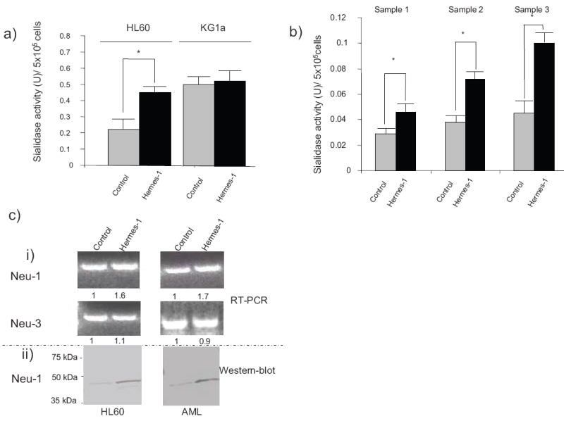
Cell surface sialidase activity was measured using 4-MU-NANA as substrate on (a) HL60 and KG1a cells, and (b) AML cells from patients (samples 1, 2 and 3), cultured in the presence or absence of Hermes-1 for 48h. Statistical significance (p≤ 0.05) for respective comparison groups is shown by brackets and asterisk. (c) (i): Representative ethidium bromide–stained gels of PCR-amplified RNA encoding sialidases Neu-1 and Neu-3 from HL60 cells and AML blasts treated with isotype-matched mAb (control) or Hermes-1 (48h treatment). Numbers indicate the relative expression of RT-PCR product normalized against GAPDH control.
(ii): Western-blot analysis of Neu-1 protein expression in HL60 cells and AML blasts treated with isotype mAb (control) or Hermes-1 (48h treatment).