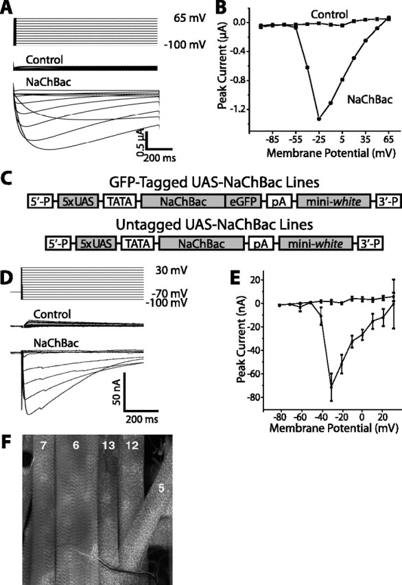
Figure 1.

Functional expression of voltage-gated bacterial sodium channel NaChBac in Xenopus laevis oocytes and transgenic Drosophila melanogaster. A, Two-electrode voltage-clamp measurements of transmembrane current in an uninjected Xenopus oocyte (control) or an oocyte injected with cRNA encoding NaChBac (NaChBac). Oocytes were held at –100 mV and stepped in increments of 15 mV to a maximum of 65 mV. Inward NaChBac currents exhibit slower activation and inactivation kinetics than those of Drosophila sodium channels that underlie neuronal action potentials. B, Current–voltage relationships for the currents measured in A. NaChBac begins to activate at approximately –60 mV. After reaching a peak at approximately –25 mV, the current begins to fall off as the transmembrane voltage approaches the reversal potential for sodium. The activation threshold of NaChBac is 20–25 mV lower than that of Drosophila neuronal sodium currents (Wicher et al., 2001). C, Constructs used for P-element transformation of the Drosophila germline. Multiple independent insertion lines were generated containing either NaChBac alone or NaChBac fused to eGFP downstream of the UAS promoter, thus allowing cell-specific expression driven by GAL4. D, E, GFP-tagged NaChBac was expressed in third instar Drosophila larval muscles using the 24B-GAL4 enhancer-trap line and the NaChBac4 insertion line, and currents were measured by two-electrode voltage clamp. Voltage-clamp recordings performed under conditions that isolate Na+ currents show robust voltage-gated inward currents with slow kinetics of activation and inactivation that begin to activate at approximately –50 mV, peak at approximately –30 mV, and fall off as the transmembrane voltage approaches the reversal potential for sodium, similar to the currents measured from NaChBac-expressing oocytes. Control muscle fibers, with the 24B-Gal4 driver alone, lack inward currents, consistent with the absence of Na+ channels in Drosophila muscle. F, Muscle fibers (as numbered) expressing GFP-tagged NaChBac channel are brightly fluorescent. The voltage-clamp measurements depicted were made in muscle fiber number 6. Error bars indicate SE.
