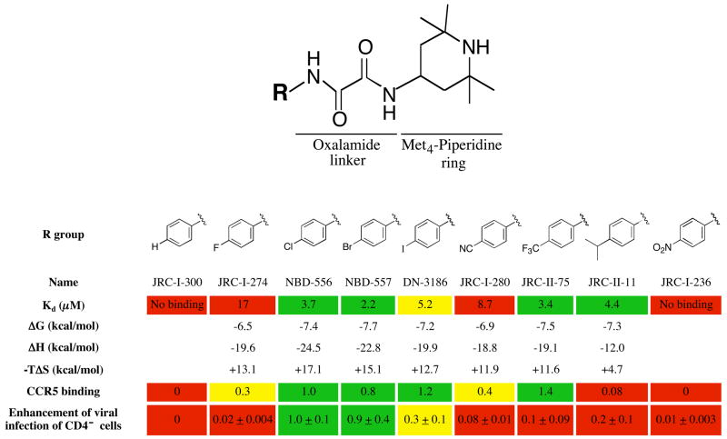
Figure 4. Structure-activity relationships of NBD-556 analogues with different para-phenyl substituents.
The values for Kd, ΔG, ΔH and −TΔS associated with the binding of each compound to the w.t. HIV-1YU2 gp120 glycoprotein were determined by isothermal titration calorimetry (see Methods). The Kd values (in μM) are color-coded as follows: red = no binding or >8; yellow = 5–8; green = <5. CCR5 binding of radiolabeled HIV-1YU2 gp120 was determined after incubation with 10 μM compound (see Methods). The induction of CCR5 binding by each compound was normalized to that observed for NBD-556. The relative induction of CCR5 binding is color-coded as follows: red = <0.25; yellow = 0.25–0.7; green = >0.7.
To study compound enhancement of infection of CD4−CCR5+ cells, recombinant, luciferase-expressing HIV-1 with the wild-type HIV-1YU2 envelope glycoproteins was incubated with increasing concentrations of the compound and then added to Cf2Th-CCR5 cells. Cells were lysed 48h later and luciferase activity measured (see methods). The area under the dose-response curve for each compound was calculated and normalized to the value obtained for NBD-556. The relative enhancement of infection of CCR5-expressing cells is color-coded as follows: red = 0–0.24; yellow = 0.25–0.7; green = >0.7.

