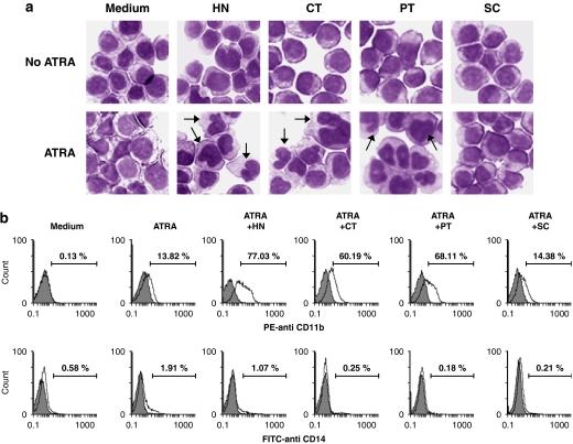
Figure 2.
Morphological and cytofluorometric analysis of four sesquiterpene lactone compounds on ATRA-induced HL-60 cell differentiation. HL-60 cells were treated for 72 h with medium alone (control), 50 nM ATRA alone or in combination with 1 μM helenalin (HT) or 10 μM of costunolide (CT), parthenolide (PT) or sclareolide (SC). (a) Cell morphology was assessed with Giemsa staining and analysed under a light microscope (magnification: × 400). The arrows indicate differentiated HL-60 cells with multilobed nuclei. (b) Cells were subjected to cytofluorometric analysis using PE-conjugated anti-CD11b mAb or FITC-conjugated anti-CD14 mAb (unshaded area) or using the isotype control mAb (shaded area). The data are representative of three independent experiments.

