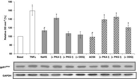
Figure 8.
Western blot analysis of NADPH oxidase in pulmonary arterial endothelial cell (PAEC) lysates using a monoclonal antibody directed against the extracellular epitope of gp91phox-subunit of mouse NADPH oxidase. Cells were either not treated or treated with tumour necrosis factor-α (TNFα) (10 ng mL−1) for 16 h with one of the following: H2S (100 nM); ACS6 (1 nM); or combination of H2S/ACS6 with protein kinase A (PKA) inhibitor (PKA I; 100 nM), protein kinase G (PKG) inhibitor (PKG I; 100 nM) or guanylyl cyclase inhibitor (ODQ; 100 nM). The middle panel shows the representative blot and the upper panel the results of the densitometric analyses of six blots (expressed as % of basal values of OD mm−2). GAPDH expression was used as a loading control (lower panel). *P<0.05; significantly increased compared with basal value. #P<0.05; significantly inhibited compared with TNFα-treated value. †P<0.01; significantly increased compared with corresponding H2S/ACS6 only values.

