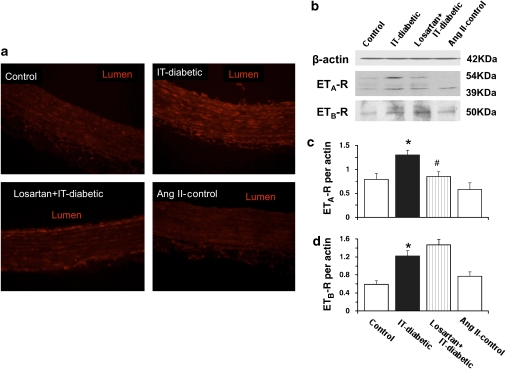
Figure 6.
Immunohistochemical staining for endothelin-1 receptor (a) and western blots for ETA receptor (ETA-R; b and c) or ETB receptor (ETB-R; b and d) in endothelium-intact rat aorta. Representative sections of endothelin-1 receptor-positive staining is shown in red for aorta obtained from control rats, with and without angiotensin II infusion and for insulin-treated diabetic rats (IT-diabetic) with and without chronic losartan therapy. Calibration bar, 25 μm. Original magnification × 100. Representative western blots of ETA and ETB receptors from rat aorta (b) were quantified by scanning densitometry (c and d). For the ETA receptor, two bands (54 and 39 kDa) corresponding to native and glycosylated forms were detected. Values are mean±s.e.mean from 8 experiments. *P<0.05 versus controls. #P<0.05 versus IT-diabetic.

