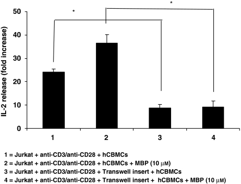
Figure 3.
Effect of cell–cell contact on Jurkat cell activation. Mast cells were co-cultured with Jurkat cells or separated by a Transwell permeable membrane (n=6). Jurkat cells were placed in the lower well and activated with anti-CD3/anti-CD28; the transwell membrane was then inserted and an equal number of mast cells were added to the upper well and activated with MBP (10 μM). After 48 h of incubation, the supernatant fluid was collected and assayed for IL-2 levels (*P<0.05 for groups compared with as shown in parentheses). MBP, myelin basic protein.

