Figure 4.

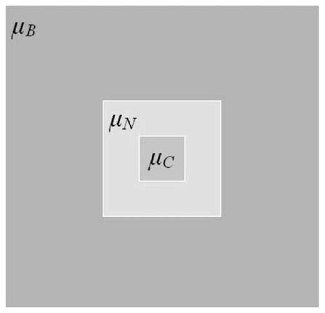
The kernel used for neighboring information. μC is the central kernel, μN is the neighboring kernel, while μB is the background kernel. This is a 2D representation of the 3D kernels.

The kernel used for neighboring information. μC is the central kernel, μN is the neighboring kernel, while μB is the background kernel. This is a 2D representation of the 3D kernels.