
Fig. 1.
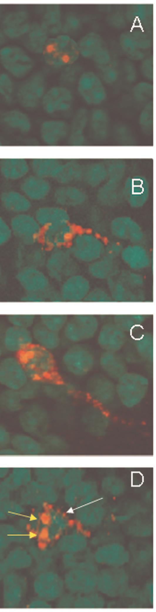
Qualitative characterization of Arc expression in granule cell neurons of the dentate gyrus. Arc was induced by 2 five minute behavioral explorations of a novel environment that were separated by 25 minutes. Intranuclear foci of Arc mRNA were induced by the second exploration, ∼ 5 minutes before tissue collection, and were detected using fluorescent in situ hybridization (A). Cytoplasmic Arc mRNA (B) or Arc protein (C) were detected in neurons activated by the first exploration. Both nuclear foci (second exploration, short yellow arrows) and cytoplasmic Arc mRNA (first exploration, long white arrow) were seen in ∼ 90% of cells immunoreactive for Arc (D). Digoxigenine labeled Arc antisense probe was detected with Cy3 (red, A, B, D), and immunofluorescence staining detected Arc protein (red, C). Cell nuclei were counterstained green and the magnification for all the images was 63×.