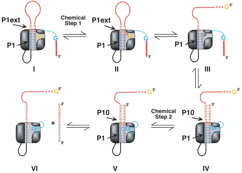
Figure 3.
Cartoon model of the self-splicing reaction. The P1 helix is shown in blue and the P1 extension and P10 are shown in red. In the first chemical step of self-splicing (transition from I to II) an exogenous G (shown in orange) attacks at the 5′-splice site. Note that in II the extended P1 duplex is nicked. In a subsequent conformational step (transition from II to III), the G, now covalently linked to the P1 extension, leaves the G binding site. Next, G414, the last nucleotide of the intron (shown in cyan), enters the G binding site and the 3′-exon is aligned in P10 for ligation (III to IV). In the second chemical step (IV to V) exons are ligated to be released in a last step (V to VI).

