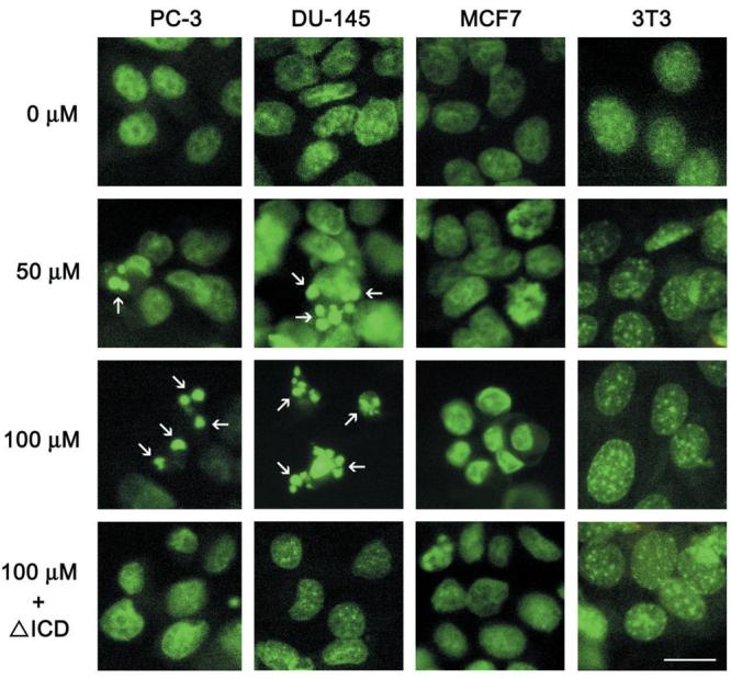
Figure 4.

Detection of apoptotic nuclei (arrows) by Hoechst staining of PC-3 and DU-145 prostate cancer cells, MCF-7 breast cancer cells and 3T3 fibroblasts following treatment with 0, 50 or 100 μM carprofen for 48 hr. Prior to treatment, some cells were co-transfected with a ponasterone A-inducible ecdysone receptor plasmid pVgRxR and ΔICDp75NTR (ICD). Following transfection, cells were incubated in serum containing medium for 18 hours, and then incubated in 1 μM ponasterone A (P) for 24 hours to drive expression of the dominant negative gene products and then treated with 100 μM carprofen for 48 hr. Scale bar = 5 μM
