
Figure 2.
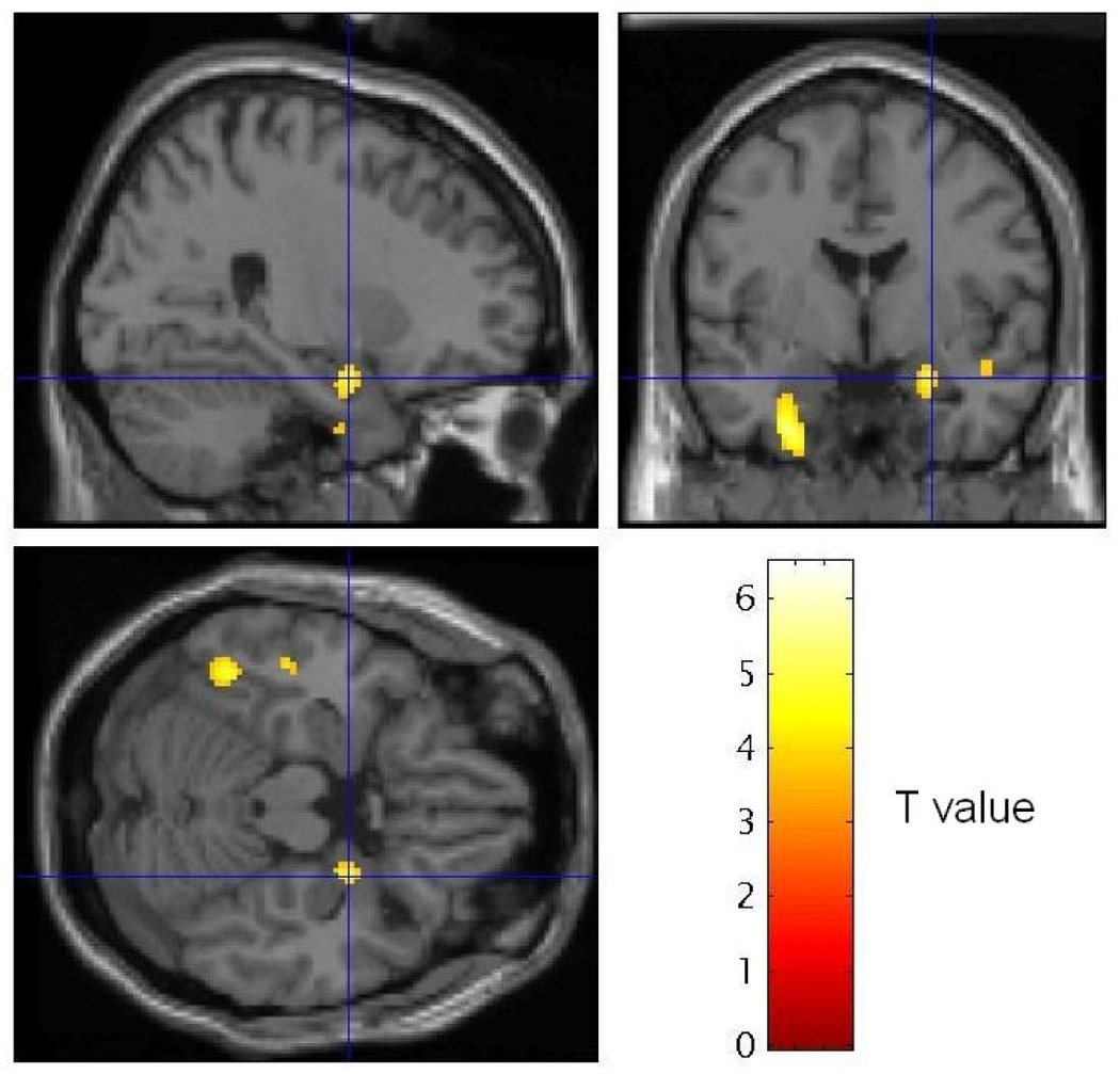
Statistical Parameteric Mapping (SPM) image sections showing voxels where normalized rCBF decreased in MVC subjects in the personalized trauma relative to neutral scripts, thresholded at T = 3.53 (p < .001). Sagittal and coronal (top row) and transverse (bottom row) sections taken at the coordinate of maximally significant deactivation in the right amygdala [26, −3, −17] are shown (coordinates shown by crosshairs are interpreted as in Table 3). An extended area of decreased blood flow also is evident in the left medial temporal cortex, located outside our a priori regions of interest. The implications of this observation will be pursued in future studies.