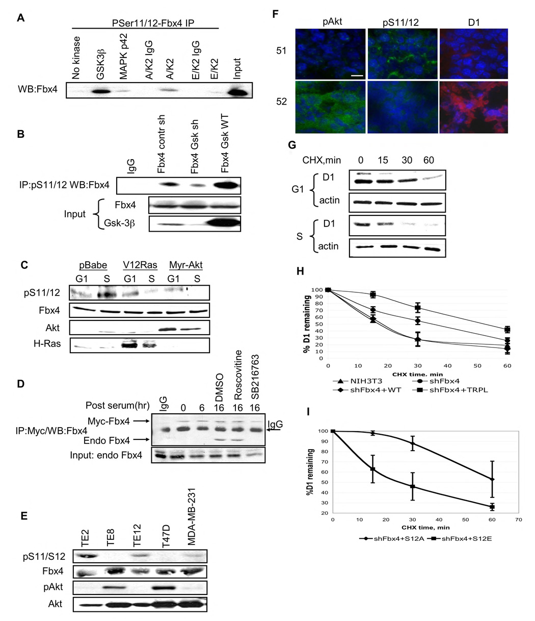
Figure 3. GSK3β phosphorylates S11/12 of Fbx4 in S phase of cell cycle.
A. In vitro kinase assay was performed using purified GST-tagged Fbx4 and indicated kinases. Following in vitro phosphorylation, samples were precipitated with the pS11/12–Fbx4 antibody and subjected to immunoblot with a Fbx4 antibody. B. 293T cells were transfected with wild type Fbx4 along with control shGFP vector, a GSK3β shRNA vector, or a plasmid encoding wild type GSK3β Fbx4 was immunoprecipitated from cellular extracts using pS11/12-Fbx4 antibody 48hrs post–transfection and detected by immunoblot with the total Fbx4 antibody. C. NIH3T3 cells were transfected with an empty vector, or RasV12 or Myr Akt expressing constructs. Cells were synchronized by serum starvation and released into complete medium for 6hrs (G1) or 16hrs(S). Protein extracts were processed for immunoblot using pS11/12-Fbx4, total Fbx4, total Akt and H-Ras antibodies. D. NIH3T3 cells were transfected with vector encoding Myc-tagged Fbx4 and 24 hrs later were synchronized in G0. Cells were released into cell cycle for the indicated intervals (cells were treated with kinase inhibitors for 6 hrs before harvesting samples at 16 hrs time point). Myc-Fbx4 pull down was performed using Myc-agarose. Myc-Fbx4 associated endogenous Fbx4 was detected by immunoblot with total Fbx4 antibody. E. Total cell extracts from esophageal carcinoma cell lines (TE2, TE8 and TE12) and breast carcinoma cell lines (T47D and MDA-MB-231) were subjected to precipitaion with the pS11/12 antibody followed by immunoblot with the Fbx4 antibody. Direct Western blotting was performed using pAkt (Ser473) and total Akt antibodies. F. Frozen sections from MMTV-Neu breast tumors were analyzed by IHC using following antibodies: pS11/12-Fbx4, pAkt (Ser473) and cyclin D1 (bar, 8µM). G. NIH3T3 cells were arrested by serum starvation following by replating in complete media. Cycloheximide was added for the indicated intervals during G1 (6 hrs post-release) and S phases of cell cycle (16 hrs post-release). Cyclin D1 turnover was analyzed by Western analysis using cyclin D1 antibody. H. NIH3T3 shFbx4 stable cell line was transfected with either wild type or Fbx4 trpl. Cycloheximide chase was performed in asynchronous cells 48 hrs after transfection. Quantification of 3 independent experiments is provided, error bars represent +/− SD. I. Fbx4S12A or Fbx4S12E were individually re-introduced into Fbx4 knockdown cells and cyclin D1 turnover was assessed in S phase cells; quantification of 3 independent experiments is provided, error bars represent +/− SD.

