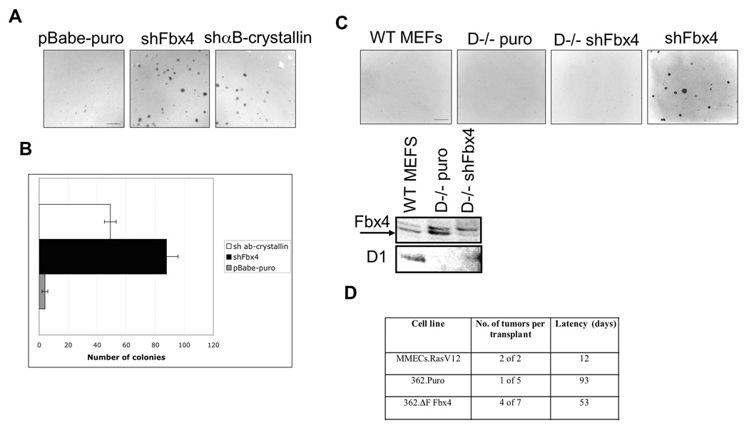
Figure 4. Inhibition of SCFFbx4/αB-crystallin activity leads to neoplastic growth.
A. Soft agar colony formation assay was performed using pBabe-puro, shFbx4 and shαB-crystallin knockdown cell lines. Cells were plated in soft agar and grown for 21 days (bar, 250µM). B. Quantification of A, error bars represent +/− SD. C. Same as A using wt and cyclin D1−/− MEFs infected with control (puro) or shFbx4 retrovirus (bar, 250µM). Levels of Fbx4 and cyclin D1 proteins are provided in the bottom panel. D. 106 cells were transplanted into cleared mammary fat pads of 3 week-old NOD-SCID mice. Mice were monitored for palpable tumor formation bi-weekly.

