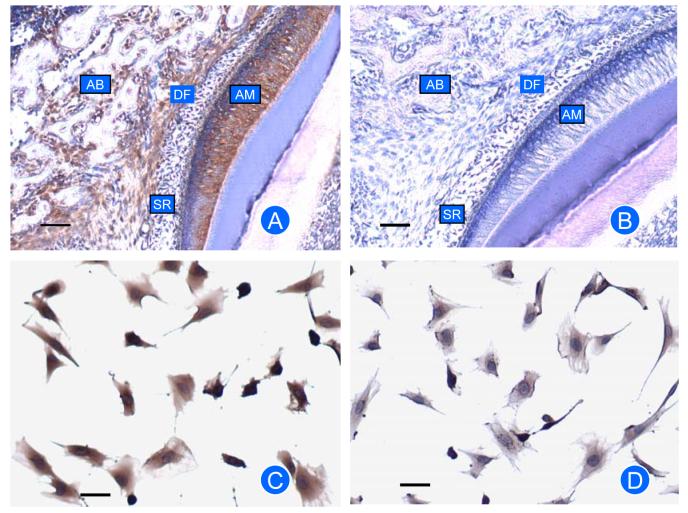
Fig. 2.
Immunostaining for EMAP-II protein in rat mandibles (A, B) and DF cells (C, D). Expression of EMAP-II was seen in dental follicle (DF) and DF cells. There is some expression in ameloblasts (AM) and alveolar bone (AB), but no expression in stellate reticulum (SR). Controls for immunostaining (B and D) with IgG used to replace the primary antibody showed little or no staining. Scale bar: 50 μm.

