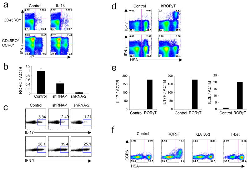
Figure 1. RORγT is necessary and sufficient for the expression of IL-17 in human CD4+ T cells.
(a) Flow cytometry on sorted CD45RO− and CD45RO+CCR6+ activated and expanded in the presence of IL-2 with or without IL-1β. IL-17 and IFN-γ production was analyzed at day 6. (b,c) RT-PCR for RORC and ACTB mRNA expression (b) and flow cytometry for intracellular IL-17 and IFN-γ (c) in sorted CD45RO+CCR6+ cells transduced with an empty vector or vector encoding for RORγT-specific shRNA (shRNA-1 and shRNA-2). Cells were selected in puromycin at day 2 and expression of mRNA and cytokines was analyzed at day 6. Data are representative of four independent experiments. (d) Flow cytometry of naive cord blood CD4+ T cells activated, transduced by vectors encoding IRES-HSA or RORγT-IRES-HSA and then expanded for 6 days in the presence of IL-2. Intracellular IL-17 and IFN-γ production was analyzed at day 6. (e) RT-PCR of ACTB, IL17, IL17F and IL26 in naive cord blood CD4+ T cells transduced with vectors encoding IRES-GFP or RORγT-IRES-GFP. GFP+ cells were sorted at day 6 and the levels of mRNAs were analyzed. (f) Flow cytometry of naive cord blood CD4+ T cells transduced with vectors encoding IRES-HSA, RORγT-IRES-HSA, GATA-3-IRES-HSA or T-bet-IRES-HSA. CCR6 cell surface expression was measured at day 12. Each panel is representative of three independent experiments unless noted otherwise.

