
Figure 3.
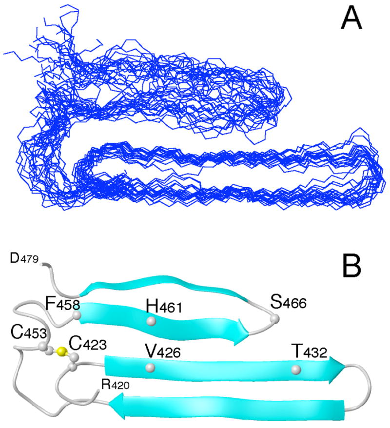
Overlay of the 20 lowest energy conformations of the bovine fibrinogen Aα406-483 αC-domain fragment (panel A). Molecules were superimposed over residues 420–478 using the backbone amide atoms C, CA, N, and O. Panel B displays the ribbon diagram of the average minimized conformation. For clarity, only the ordered residues, 420–478, are shown. The locations of some residues mentioned in the text, including C423 and C453 forming the disulfide bond, are indicated.