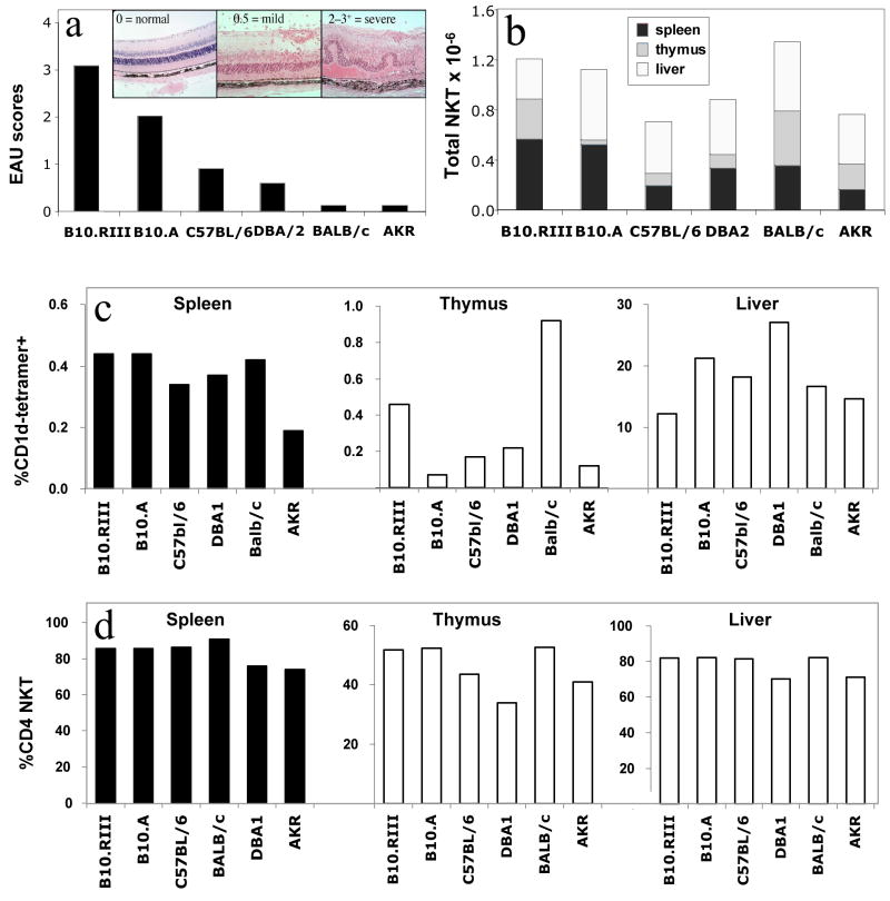
Fig. 1. Total iNKT cell numbers and percentages of CD4+ iNKT cells seem unrelated to susceptibility to EAU.
Six different mouse strains (six mice per strain, 8 weeks of age) were immunized with 150 μg IRBP in Complete Freund’s Adjuvant and an additional i.p. injection of 0.3 μg Pertussis Toxin (PTx). B10.RIII mice were immunized with 10 μg IRBP without additional PTx. (a): Schematic representation of typical strain-specific disease scores and representative histopathology (inset), based on established data. Lymphocytes from naïve livers, thymuses and spleens were isolated on a density gradient and iNKT cell numbers were determined by flow cytometry using labeled CD1d/α–GalCer tetramers, anti-CD4 and anti-β-TCR antibodies as described in Materials &Methods. (b): Average of isolated iNKT cell numbers in thymus, LN and spleen, expressed as a total cell number collected from each mouse. (c): Average of isolated iNKT cell numbers in thymus, LN and spleen, expressed as a percentage of the total cell number collected from each mouse. (d): Proportion of CD4+ iNKT cells expressed as a percentage of total iNKT cells. The data are pooled from 2 identical experiments of 3 mice each (total 6 individual animals per point).

