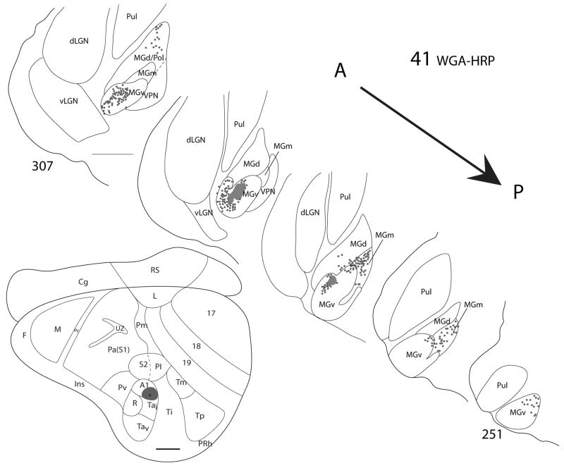
Figure 6.
Thalamic connections of A1 in squirrel 41. The WGA-HRP injection was guided by microelectrode mapping. The injection was mostly within A1 but extended slightly into the cortex ventral to A1 (Tai). The location of the injection site is indicated on a representative drawing of the grey squirrel flattened cortex. Thalamic sections are arranged in an anterior (section 307) to posterior (section 251) progression. Dots indicate the locations of retrogradely labeled cell bodies whereas patches represent the locations of possible anterograde label. Labeled cells are primarily concentrated in MGv and also present in MGm and MGd. Scale bar for brain = 2mm, for thalamic sections = 1mm.

