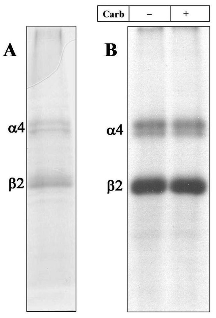
Figure 1. Photoincorporation of [125I]TID into Purified α4β2 nAChR.
An aliquot of affinity-purified α4β2 receptor (50 µg) was equilibrated for 1 h with [125I]TID (0.4 µM; 10 µCi), in the absence (− lanes) and in the presence (+ lanes) of 400 µM carbamylcholine (Carb) then irradiated at 365 nm for 7 min. The protein was pelleted by centrifugation, resuspended in electrophoresis sample buffer and fractionated by SDS-PAGE. Following electrophoresis, the mapping gel was stained with Coomassie Blue R-250, destained, dried and exposed to X-ray film with an intensifying screen. A, Coomassie stained gel. B, Autoradiograph, 15 h exposure. The electrophoretic mobility of α4β2 receptor subunits are indicated on the left.

