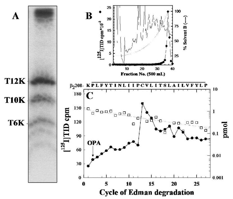
Figure 5. [125I]TID labels β2-Cys220 in β2M1.
The β2V8-21 fragment, produced by in-gel digestion of [125I]TID-labeled β2 subunit with V8 protease (Figure 2B), was digested with trypsin for 5 days and the digest was fractionated on Tricine SDS-PAGE. A, Autoradiograph of a Tricine gel showing [125I]TID photoincorporation into three peptide fragments with apparent molecular masses of 12 KDa (β2T12K), 10 KDa (β2T10K) and 6 KDa (β2T6K). B, Reversed-phase HPLC purification of [125I]TID-labeled β2T12K. C, 125I (●) and PTH-amino acids released (□) during sequencing of β2T12K HPLC fractions 36–38, with sequencing interrupted for OPA treatment before cycle 2. The primary amino acid sequence began at β2-Lys208 ( Io, 1.2 pmol; R, 93 %; 18,630 cpm loaded in the filter and 11,909 cpm left after 30 cycles) and the peak of 125I release was in cycle 13 (90 cpm) correspond to labeling of β2-Cys220. The amino acid sequence detected is shown above the panel, with a line indicating the M1 region.

