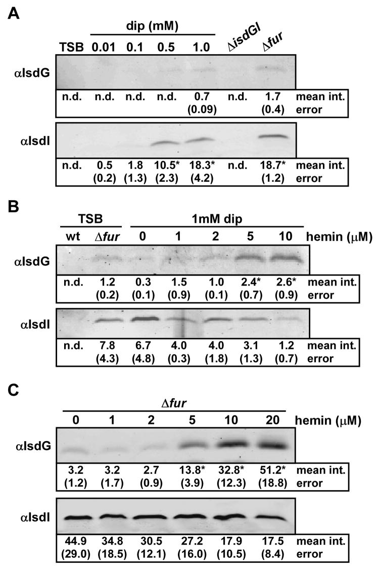
Figure 2. Regulation of IsdG and IsdI by iron and hemin.
S. aureus strains were grown overnight in the indicated media and protoplast fractions were harvested and normalized by total protein concentration. The mean intensity of each band, as determined by densitometric analyses, is presented under the blots in arbitrary units, with corresponding standard error. Bands that were not detected for quantification are reported as not determined (n.d). Statistical significance was determined using the Student’s t test. Results are representative of at least four independent experiments. (A) The effect of iron chelation by 2,2’dipyridyl (dip) on expression of IsdG and IsdI was analyzed by immunoblot. Asterisks denote statistically significant increases, as compared to 0.01 mM dip (p < 0.0001). (B) The effect of exogenous hemin on expression of IsdG and IsdI in iron-deplete media (dip) was analyzed by immunoblot. Asterisks denote statistically significant increases, as compared to 0 μM hemin (p < 0.03). The slight decrease in IsdI abundance in 10 μM hemin compared to 0 μM hemin is not statistically significant (p > 0.06). (C) The effect of exogenous hemin on expression of IsdG and IsdI in an isogenic Δfur strain was analyzed by immunoblot. Asterisks denote statistically significant increases, as compared to 0 μM hemin (p < 0.0002). The slight decrease in IsdI abundance in 20 μM hemin compared to 0 μM hemin is not statistically significant (p > 0.1).

