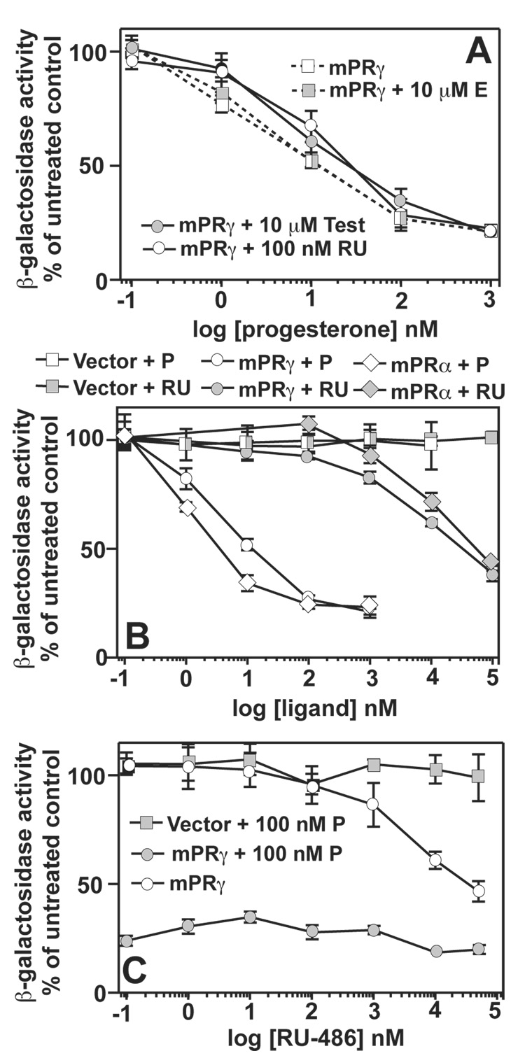
Figure 2. Steroid specificities for mPRγ and mPRα.
In all cases, FET3 expression is measured using the FET3-lacZ reporter. All PAQR are cloned into the pGREG536 vector. All cells are wild type and are grown in iron-deficient LIM containing 0.05% galactose/1.95% raffinose. (A) The dose response of FET3 in cells expressing mPRγ plasmid to progesterone either alone or in the presence of 10 µM β-estradiol (E), 10 µM testosterone (test) or 100 nM RU-486 (RU). (B) Dose response of FET3 to either progesterone (P) or RU-486 (RU) in cells carrying either empty expression vector or vectors that express mPRγ or mPRα. (C) Dose response of FET3 to RU-486 in cells carrying either empty expression vector or a vector that expresses mPRγ. Cells are either treated with RU-486 alone or in the presence of 100 nM progesterone (P). When data points overlap or are very close to overlapping, combined symbols are used.

