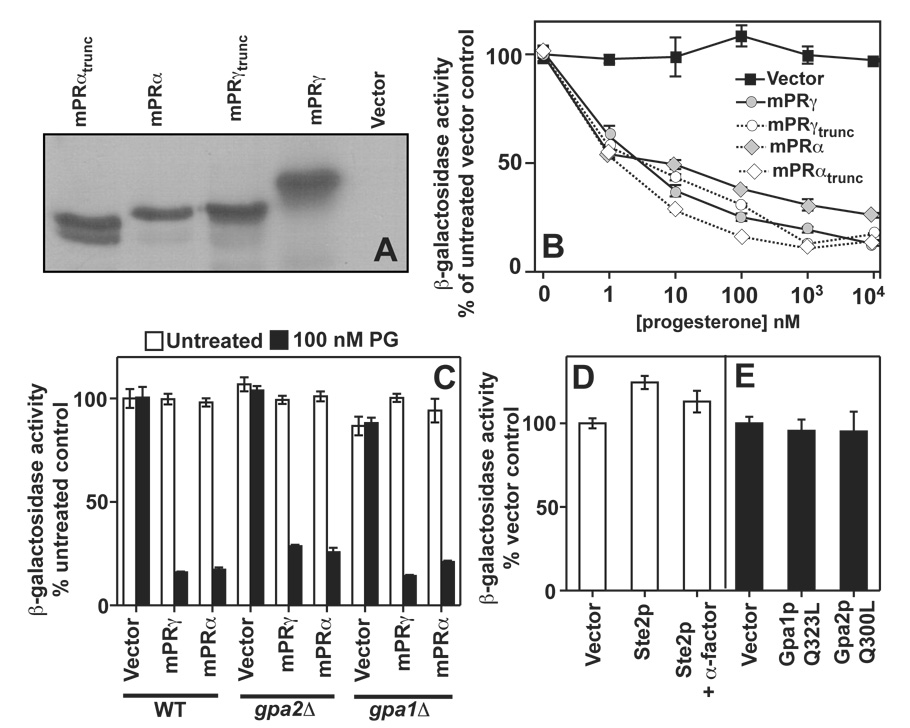
Figure 6. Truncation mutations and G-protein signaling.
In all cases, FET3 expression is measured using the FET3-lacZ reporter. (A) Full length and truncated mPRγ and mPRα were expressed in wild type cells grown in 2% galactose from the pGREG536 vector. The location of the C-terminal truncations is shown in Figure 4. All proteins possess an N-terminal 7x-HA tag. Proteins were detected by Western blot using an anti-HA antibody. (B) The dose response of FET3 to progesterone in cells expressing the full length or truncated mPRγ and mPRα and grown in 0.05% galactose/1.95% raffinose. (C) The ability of mPRα and mPRγ to respond to progesterone and repress FET3 is not impaired in either gpa2Δ or gpa1Δ cells (gpa1Δ cells also lack the STE7 gene, see text) (D) Overexpression of the Ste2p GPCR using the GAL1 promoter does not repress FET3 in wild type cells (mat a) grown in 2% galactose. Activation of overexpressed Ste2p via the addition of 1 µM of its agonist, α-factor, also has no effect on FET3 under these conditions. (C) Expression of constitutively active alleles of Gpa1p (Gpa1pQ323L) and Gpa2p (Gpa2pQ300L) from the GAL1 promoter had no effect on FET3 in cells grown in 2% galactose.

