
Figure 2.
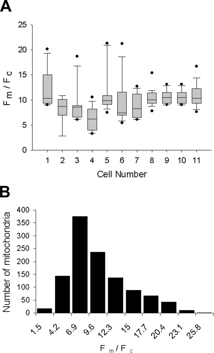
Samples of Fm/Fc show greater intercellular than intracellular variation. A, The mitochondrial fluorescence ratio distribution is shown for a representative sample of 11 neurons using a box plot and shows the mean (middle hash mark), 25th and 75th percentiles (top and bottom of box), 10th and 90th percentiles (top and bottom error bars), and any data points beyond the 10th and 90th percentiles (top and bottom dots). B, Histogram of Fm/Fc shows a larger sample of 94 neurons with 1122 mitochondria but includes the sample of 11 neurons shown in A. The total population of Fm/Fc does not show a normal distribution but has a tail with a higher Fm/Fc. All mitochondria were sampled from the axon shaft with 5–15 mitochondria per axon.