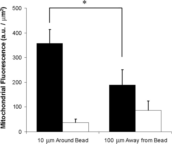
Figure 6.
Mitochondria accumulate at sites of contact between the axon and semaphorin 3A-coated beads. The total TMRM fluorescence was compared between regions around either sema3A-coupled beads (black bars; n = 28 cells) or heat-treated inactive beads (white bars; n = 20 cells). The 10 μm immediately surrounding the bead was compared with a 10 μm region of the axon 100 μm away from the bead to be far enough away from any effect from the bead. Mitochondria were often closely adjacent to one another and could not be distinguished individually, so total fluorescence was compared between the two regions. Mitochondria accumulate around the sema3A beads shown by a significant increase in mitochondrial fluorescence when compared with heat-inactivated beads by a t test. *p < 0.001. Error bars show SEM.

