
Figure 7.
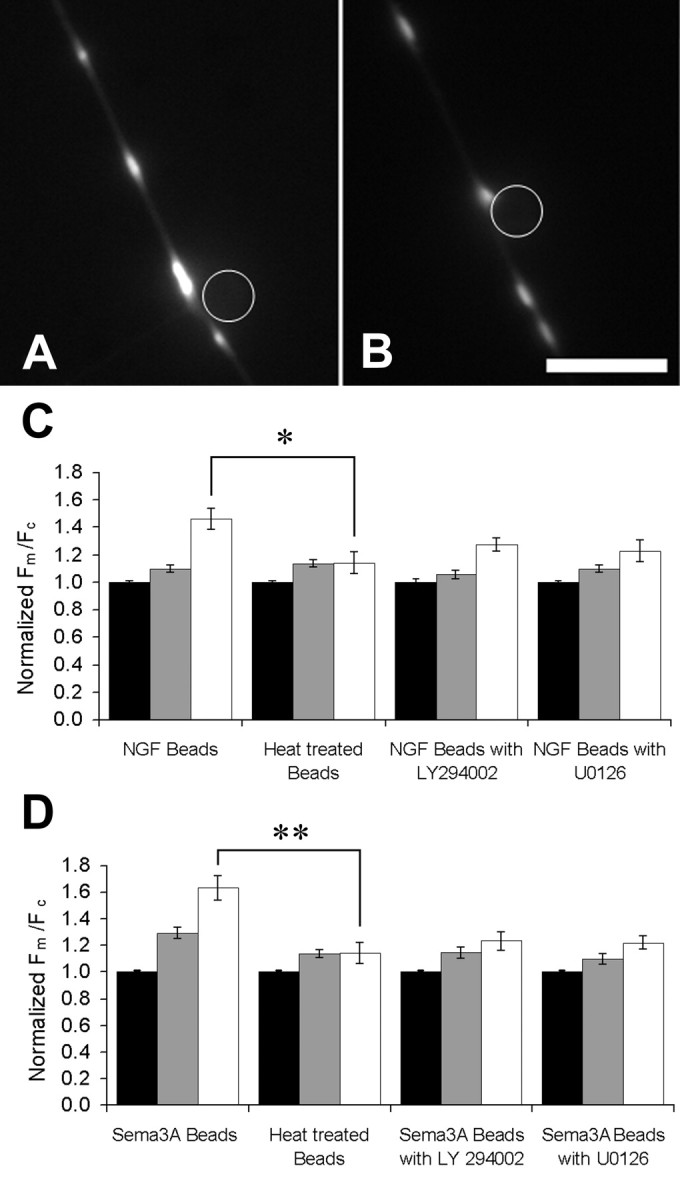
Axons incubated with NGF- or semaphorin 3A-coated beads display a local increase in Fm/Fc in the region of axon immediately adjacent to the bead. Cells were rinsed and incubated in very low NGF medium and beads for 1 h before experiments. Fluorescence images show sites where an NGF-coated bead (A) and a sema3A-coated bead (B) are in contact with otherwise undistinguished regions of axons. Scale bar, 10 μm. C, The distance between the mitochondria and the bead was measured and used to sort them into three groups: those in the 10 μm of axon centered on the bead (white bars), those between 5 and 50 μm away (gray bars), and those >50 μm from the bead (black bars). The mean Fm/Fc of axonal mitochondria was increased within 50 μm of the site of contact with an NGF bead, and this effect was even greater for mitochondria in the 10 μm region centered on the contact site. The increase in Fm/Fc near the NGF bead was partially blocked by either PI3 kinase inhibitor LY294002 or MAPK inhibitor U0126 for 20 min. From left to right, n = 18, 22, 20, and 16 neurons. D, The mitochondria in axons treated with sema3A-coated bead were sorted according to distance from the site of axon–bead contact as just described. The mean Fm/Fc of axonal mitochondria was also increased within 50 μm of the site of contact with a sema3A bead, and this effect was even greater for mitochondria adjacent to the contact site. The increase in Fm/Fc near the sema3A bead was partially blocked by PI3 kinase inhibitor LY294002 or MAPK inhibitor U0126. Heat denaturation of both kinds of beads eliminated their effect on Fm/Fc. From left to right, n = 18, 17, 21, and 16 neurons. *p < 0.05; **p < 0.01. Error bars show SEM.