
Figure 3. Peripheral Branches of Trigeminal System II.
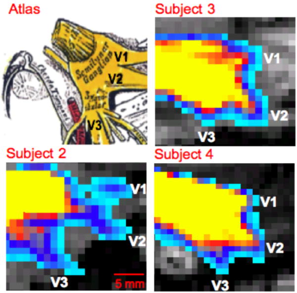
Trigeminal nerve branches at the level of the TG are shown in three other subjects (See also Segmentation 1 in Methods and Materials). In each case the seeding masks for probabilistic tractography was placed in the TNR or TG. All possible pathways through the seed regions were identified (waypoints not implemented). An atlas based figure is shown in the upper left corner (25).