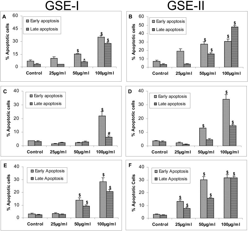
FIG. 5.
GSE-I and GSE-II preparations induce apoptotic cell death in a panel of human CRC cell lines. LoVo, HT29 and SW480 cells were plated and treated with DMSO (control) or varying concentrations of GSE-I or GSE-II (25-100μg/ml) as detailed in the Materials and Methods. Following 24h of these treatments, adherent and non-adherent cells were collected by brief trypsinization and processed for FACS analysis following annexin V-PI staining. Panels A and B represent data for GSE-I and GSE-II treatment of LoVo cells; panels C and D represent data for GSE-I and GSE-II treatment of HT29 cells; and panels E and F represent data for GSE-I and GSE-II treatment of SW480 cells. The data shown are mean ± SD of three independent plates. *, P < 0.05; #, P < 0.01; and $, P < 0.001, control (DMSO) versus various GSE-I/GSE-II treatments as indicated in the figure.

