Figure 2.
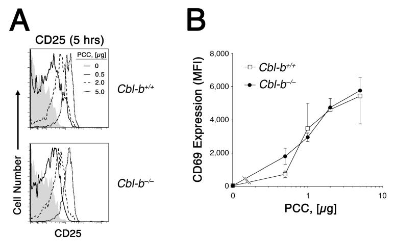
Cbl-b does not raise the threshold for initial Ag recognition. 2 × 10⁶ CFSE-labeled Rag2-/- 5C.C7 CD4+ T cells were adoptively transferred into B10.A recipients and exposed to varying doses of PCCp. 5 h later, animals were sacrificed and CFSE-labelled lymph node CD4+ T cells were examined for expression of activation markers. A, CD25 expression by T cells from wildtype (top) and Cbl-b-deficient (bottom) donor mice exposed to either 0 μg (shaded histogram), 0.5 μg (solid tracing), 2.0 μg (dashed tracing), or 5 μg (dotted tracing) PCCp infusion. B, CD69 expression by wildtype (open square) and Cbl-b-deficient (closed circle) CD4+ T cells following immunization with the indicated dose of PCCp. Two to three animals were examined at each dose of Ag, and data are expressed as the mean fluorescence intensity ± SEM.

