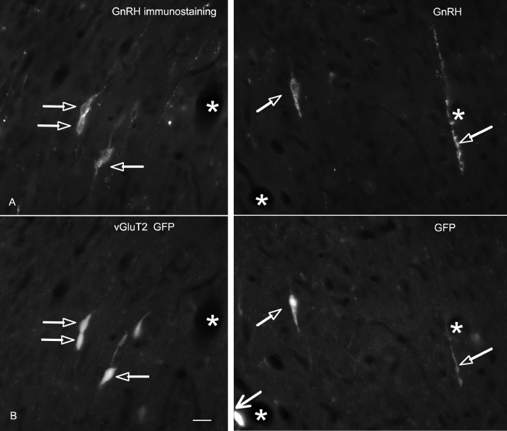
Figure 5.
GnRH immunoreactivity colocalizes in vGluT2-GFP neurons. A, GnRH-immunoreactive perikarya and processes in the MSDB of vGluT2-GFP mice. B, vGluT2-GFP-immunoreactive perikarya and processes. Note the double-labeled structures (open arrows). Also note the strongly immunoreactive vGluT2-GFP neuron in the bottom of the field (larger arrow) that was not GnRH positive. Thus, some but not all vGluT2-GFP neurons colocalize GnRH immunoreactivity. Asterisks indicate blood vessels.

