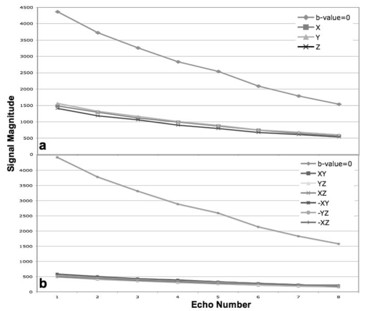
FIG 4.
Plots of the average echo magnitude data collected from the gel phantom with the modified radial-FSE sequence containing mixed-CPMG phase cycling and using a refocusing slice 300% wider than the excitation slice. The individual diffusion gradients with b-value = 500 s/mm2 is shown in a. Six DTI directions with b-value = 1000 s/mm2 are shown in b. Notice the smooth, non-oscillatory, decay along the echo train for all diffusion directions.

