
Fig. 4.
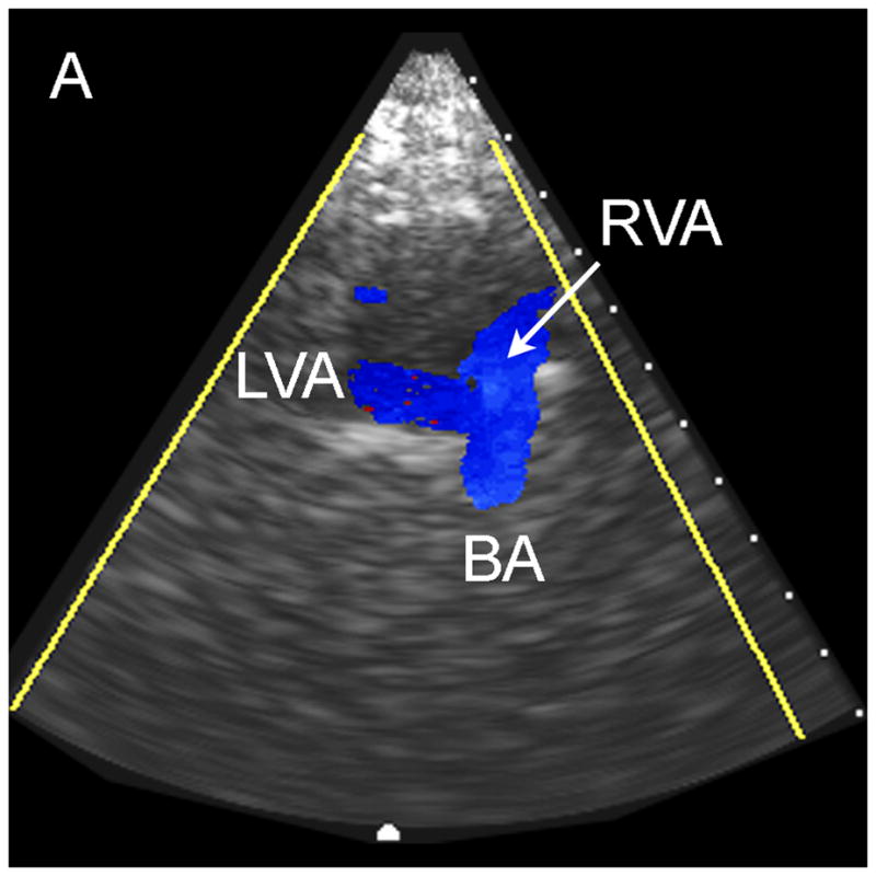
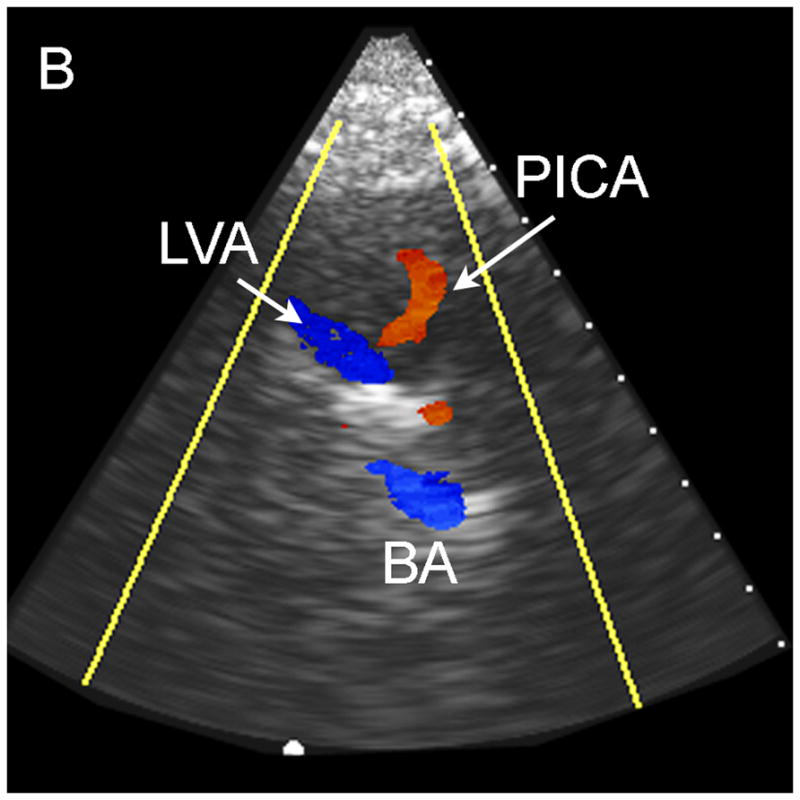
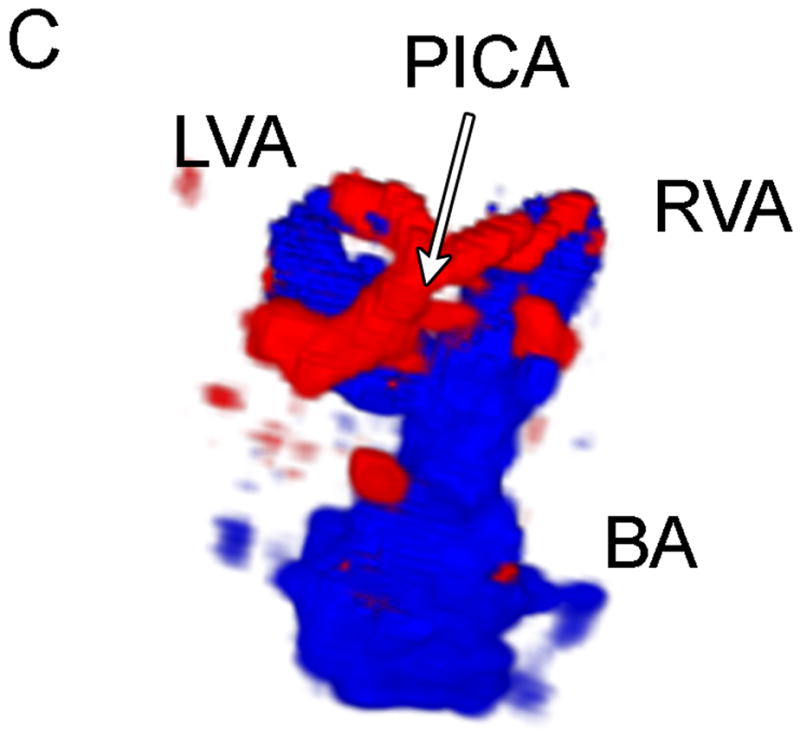

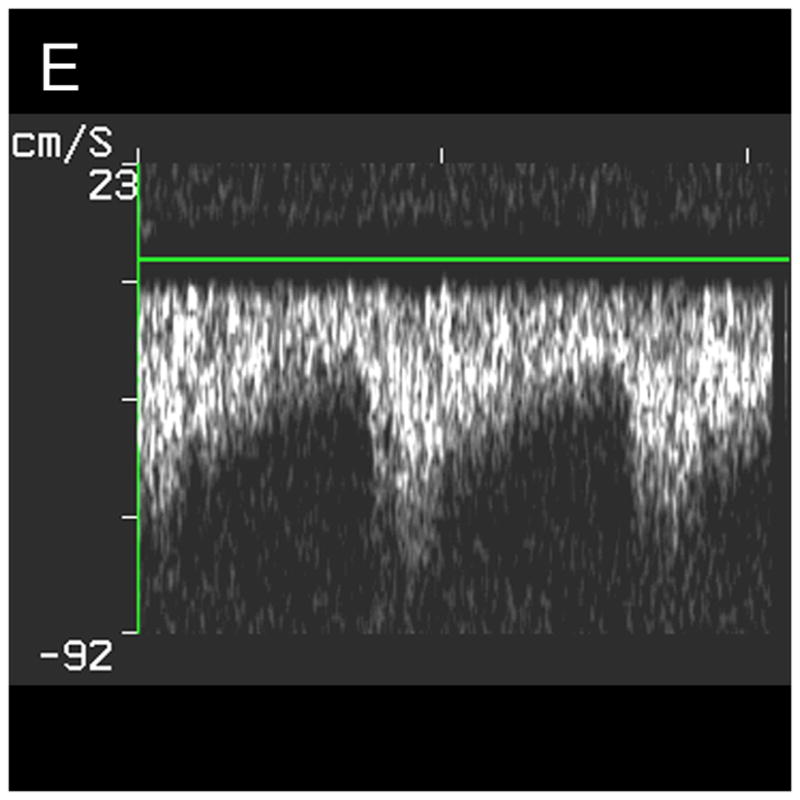
Typical Sub-Occipital Window Findings. (a) Sub-occipital coronal scan showing the junction of the left and right vertebral arteries (LVA and RVA, respectively) into the basilar artery (BA). (b) Sub-occipital parasaggital scan showing the PICA bifurcating from the LVA. (c) 3D Doppler rendering of vertebrobasilar circulation. (d) Schematic of (c). (e) Spectral Doppler trace from a BA showing velocity over the full cardiac cycle.