
Figure 1. Th17 Cells Are Preferentially Present in the Small Intestine.
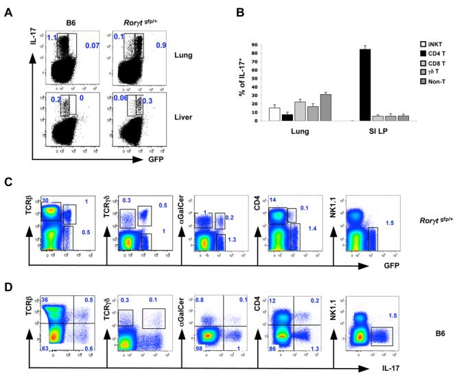
(A) Expression of GFP and IL-17 in total cells from lung and liver of Rorγt gfp/+ and C57BL/6 mice. Data are representative of 3 independent experiments. Lung and liver cells were isolated as described in Methods. All plots were gated on lymphocytes.
(B) Representation of different cell populations among the IL-17+ cell fraction in lungs and small intestines (SI LP) of C57BL/6 mice. Data are combined from three independent experiments. N = 5 mice for lung and N = 8 mice for the SI LP. Error bars represent standard deviation of the mean.
(C, D) Expression of GFP (C) and IL-17 (D) in total cells from lung of Rorγt gfp/+ (C) and C57BL/6 (D) mice. Data are representative of 3 independent experiments. Lung cells were isolated as described in Methods. All plots are gated on lymphocytes.