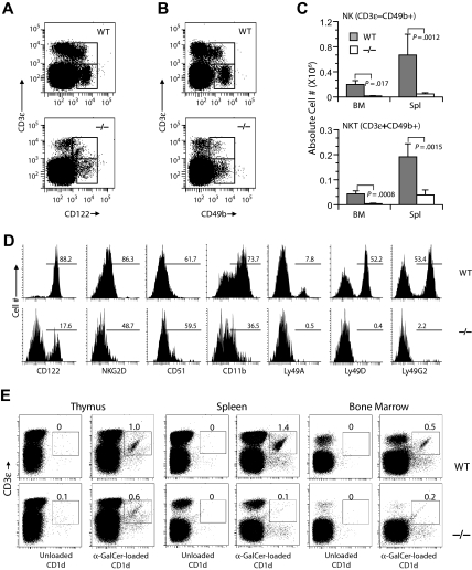
Figure 5.
Gimap5−/− mice lack NK and NKT cells. Development of NK and NKT cells is severely impaired in Gimap5−/− mice. (A) Representative analysis of BM from Gimap5−/− and WT mice stained with anti–CD3-ϵ and anti-CD122; numbers indicate percentage of cells (average ± SD) in gate. CD3-ϵ−CD122+ NK and CD3-ϵ+CD122+ NKT are significantly reduced in Gimap5−/− mice (NK: P = .004; NK-T: P = .002). (B) Representative analysis of BM from Gimap5−/− and WT mice stained with anti–CD3-ϵ and CD49b. (C) The absolute numbers (average ± SD) of CD3-ϵ−CD49b+ NK or CD3-ϵ+CD49b+ NKT cells were significantly reduced in the Gimap5−/− mice. Absolute cell numbers were calculated as percentage cells times percentage lymphocyte times cellularity of spleen (Spl) or BM (n = 6/group). P values calculated by the Student t test. (D) Expression of developmental markers in BM-derived fresh Gimap5−/− CD3-ϵ−CD49b+ NK cells is severely reduced. The frequencies of cells positive for each marker among CD3-ϵ−CD49b+ NK cells are shown along with representative histograms. Gates were set using unstained or nonspecific isotype antibody controls (not shown). Data are representative of 4 mice/genotype. (E) Gimap5−/− mice lack CD1-αGalCer+ NKT cells. Single-cell suspensions prepared from the indicated tissues of wild-type mice (WT) and Gimap5−/− mice (−/−) were stained with anti–CD3-ϵ mAb and α-GalCer–loaded CD1d dimers. Numbers represent percentage Vα14+ CD3-ϵ+ iNKT cells in the gates shown. Anti–CD3-ϵ mAb and unloaded CD1d dimers were used as background controls. Data presented are representative of at least 3 independent analyses.

