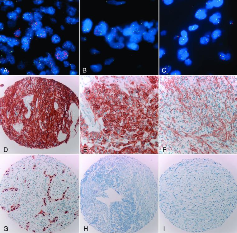
Figure 4.
Abnormalities of EGFR and cell cycle inhibitors. Dual color FISH studies (A-C) and immunohistochemical stain for EGFR (D-F). EGFR amplification and overexpression were frequent in E-GBM (A,D). EGFR amplification was a focal finding in one case of TE-GBM, being present in the adenoid/epithelial (B) but not the glial (C) component. Immunohistochemistry was patchy in this case, with strong overexpression of EGFR in some “adenoid/epithelial” fields (E) but not in others (F). Epithelial component of a TE-GBM demonstrating strong immunoreactivity for p53 (G). Loss of p21 immunoreactivity in the epithelial and glial components of a TE-GBM (H). Loss of p27 expression in the glial and sarcomatous components of a gliosarcoma (I).

