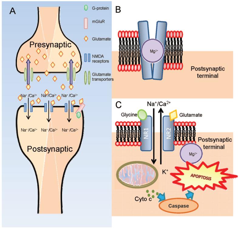
FIGURE 2.
A, Upon excitation by the nerve impulse, glutamate is released from the presynaptic terminal into the synaptic cleft where it binds onto the NMDA-receptors located on the postsynaptic terminal. Glutamate transporters located presynaptic terminal actively transport glutamate back into the terminal. B, Glutamate and glycine bind onto the receptors and the interaction causes transient conformational change in the channel and the depolarization of the cell resulting in the liberation of the Mg2+ which blocks the channel. The opening of the channel allows extracellular ionic molecules such as Ca2+ and Na+ to diffuse through the channel into the cell. Under normal physiological conditions, the NMDA channel is closed and is open transiently to enable the generation of a nerve impulse. C, When there is excessive glutamate present, the channel remains open causing a flood of Ca2+ and Na+ resulting in the depolarization of the mitochondrial membrane potential. Such events trigger off the release of cytochrome c which subsequently activates the caspase pathway leading toward apoptosis. A color version of this figure is available at www.optvissci.com.

