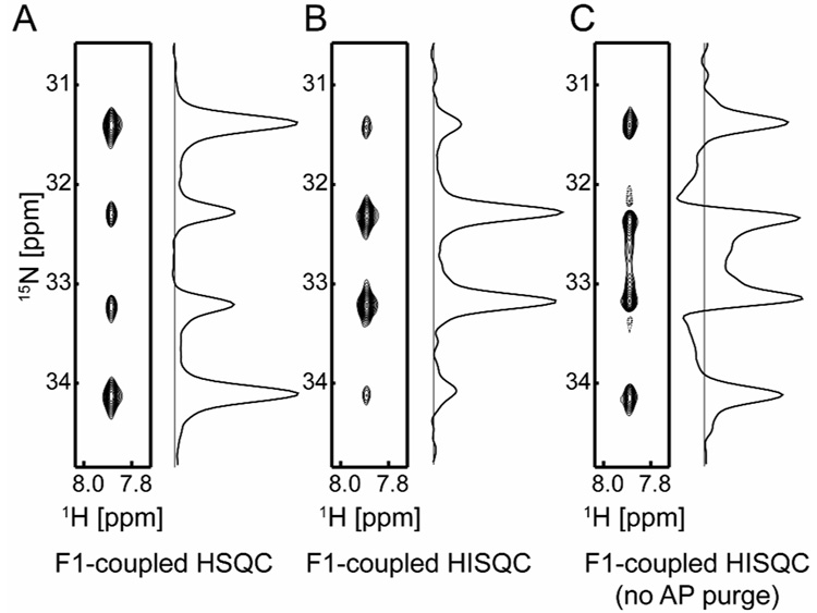
Figure 2.
15N multiplets observed for the Lys57 NH3+ group of 2H/15N-labeled homeodomain bound to 24-bp DNA (Solid contours, positive; Dashed, negative). Spectra in panels A, B and C were recorded at 16 °C with the pulse sequences shown in Figures 1A, 1B, and 1C, respectively. The 15N carrier position was at 30 ppm and r-SNOB pulses[14] selective to lysine 15Nζ nuclei were employed for 15N 180° pulses. Acquisition times for 1H and 15N dimensions were 54 ms and 79 ms, respectively. For data processing, 60°-shifted sine-bell window functions were applied prior to Fourier transformations. The protein-DNA complex was prepared as described previously [15–18] and dissolved with a buffer of 20 mM sodium phosphate and 20 mM NaCl (pH 5.8, 100% 1H2O). The solution was sealed into the inner compartment of the co-axial NMR tube, and D2O for NMR lock was put in the outer compartment to avoid NH2D and NHD2 species[2]. Data were collected at 1H-frequency of 800 MHz and analyzed with the NMRPipe[19] and NMRView[20] programs. The J-coupling was measured to be 74 Hz.

