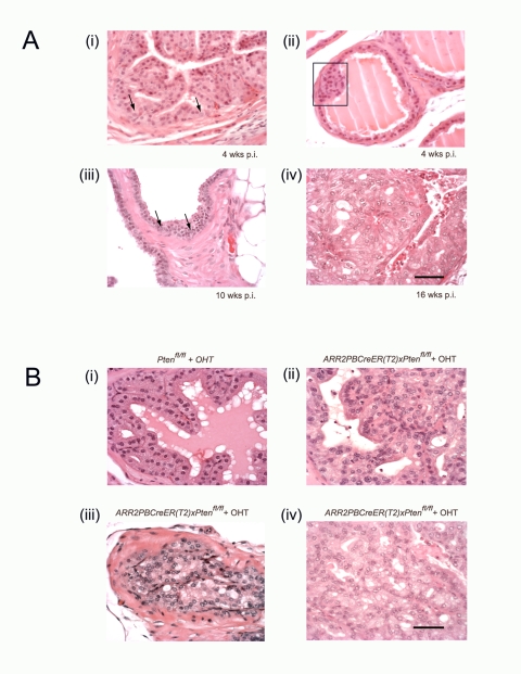
Figure 1. Transition from the earliest stages of transformation to occur progressively in ARR2PBCreER(T2)×Ptenfl/fl mice following OHT exposure.
(A) Nuclear enlargement with prominent and multiple nucleoli is seen in the luminal epithelial layer at different times post-OHT; in single cells at 4 wks post-OHT (arrows (i)); becoming more widespread at 8–10 wks post-OHT (arrows (iii)) and in the majority of cells in PIN lesions at 16–20 wks post-OHT (iv). Low grade hyperplasia is seen in the luminal layer at 4 wks post-OHT (rectangle (ii)). At 10 wks post-OHT, hyperplasia became more pronounced and nuclear enlargement with prominent nucleoli and hyperchromasia became more prevalent (arrows (iii)). Early stages of PIN, such as nuclear overlapping and mild tufting are noticeable by 10 wks post-OHT (iii). At 16–20 wks post-OHT more advanced PIN stages were evident (iv), with overt tubular dysplasia while basement membranes remained unbreached. PIN was prominent and most cells within these lesions showed nuclear enlargement, prominent nucleoli, hyperchromasia, and abnormal morphology (iv). (B) High grade PIN lesions were present in mice at 16–20 wks post-OHT. (i) While Ptenfl/fl controls showed normal morphology after OHT treatment, PIN lesions are seen 16–20 wks post-OHT administration in ARR2PBCreER(T2)×Ptenfl/fl mice, with different categories of high grade PIN being evident; including as tufting, micropapillary and flat atypia (ii), and cribriform structures (iii) and (iv) were seen in both focal intra-tubular as well as the more widespread lesions in the experimental animals. Scale bars: 50 µm.

