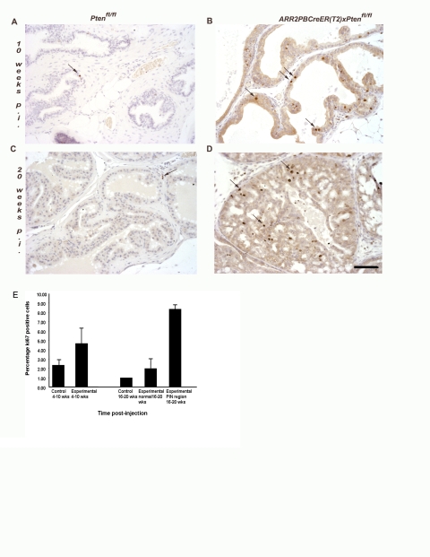
Figure 2. There is increased cell proliferation in the luminal epithelia of ARR2PBCreER(T2)×Ptenfl/fl mice exposed to OHT.
Immunohistochemical analyses revealed increased numbers of proliferating cells within the luminal epithelium of ARR2PBCreER(T2)×Ptenfl/fl mice treated with OHT at 6 wks of age as indicated by Ki67 staining, as shown by the dark dots and arrows in (B) and (D). (A) and (C) control Ptenfl/fl at 10 wks and 20 wks post-OHT; (B) ARR2PBCreER(T2)×Ptenfl/fl at 10 wks post-OHT and a (D) a representative PIN lesion in a ARR2PBCreER(T2)×Ptenfl/fl mouse at 20 wks post-OHT. Scale bar: 50 µm. (E) Quantification of the percentage of proliferating cells in control and ARR2PBCreER(T2)×Ptenfl/fl aged for 4–10 wks and 16–20 wks post-OHT injections. There is a significant increase in the percentage of proliferating cells in the PIN regions of 16–20 p.i. experimental animals both over control animals and the normal regions within the experimental animals (p-value<0.001).

