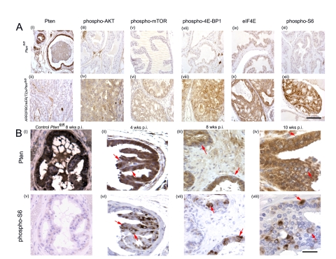
Figure 3. Downstream components of the PI3K signaling pathway are activated in PIN lesions of ARR2PBCreERT2×Ptenfl/fl mice post-OHT exposure.
(A) Immunohistochemistry showing decreased Pten levels in a representative PIN lesion ((ii) bottom panel); top panel (i) shows Pten levels in prostate epithelium of control Ptenfl/fl mice. Immunohistochemistry revealed increased levels of phospho-AKT, phospho-mTOR, phospho-4E-BP1, eIF4E, and phospho-S6 (bottom panels (iv); (vi); (viii); (x); (xii) respectively) in PIN regions of positive animals (bottom panels) as compared to controls (top panels (iii); (v); (vii); (ix); (xi)). (B) Pten loss in the prostatic epithelium is associated with increase in pS6 ribosomal protein levels in the preneoplastic phase of ARR2PBCreER(T2)×Ptenfl/fl mice post-OHT. Serial sections stained with either anti-Pten or anti-pS6 antibodies showed that loss of Pten could be correlated with increased phospho-S6 signal and that this could be detected at the single cell level during early time points post-OHT (arrows), even at a time when cellular and nuclear atypia were not present; as indicated by the red arrows - (ii) and (vi) 4 wks; (iii) and (vii) 8 wks; (iv) and (viii) 10 wks post-OHT. (i) and (v) show Pten, and phospho-S6 immunoreactivity, respectively, in serial sections form the prostate of a Ptenfl/fl control mouse 8 wks post-OHT. Scale bars: A (i–xii), 100 µm; B (i–vi), 37.5 µm.

