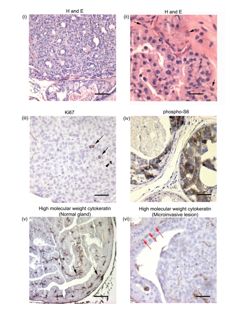
Figure 4. Transition from PIN to invasive carcinoma is seen in ARR2PBCreER(T2)×Ptenfl/fl mice exposed to OHT and aged for 42–52 wks.
(i) Widespread high grade PIN lesions were seen in multiple glands within the prostates of experimental animals at 42–52 wks post-OHT, (ii) with abnormal cellular and nuclear morphology and microinvasive lesions (arrows). (iii) Proliferating cells within these lesions were detected by immunostaining with the anti-Ki67 antibody (arrows; arrowhead shows a mitotic figure) and high levels of phospho-S6 expression were also detected (iv). Representative immunohistochemical analysis using an antibody against high molecular cytokeratins 1,5,10, and 14 that stains basal cells: although basal cytokeratin staining was present within normal-appearing glands (v) (black arrows), with progression to invasive carcinoma there was a lack of cytokeratin staining (vi) (red arrows) within the same prostate. Scale bars: (i), 100 µm; (ii–vi), 25 µm.

