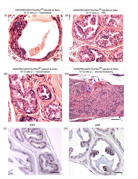
Figure 5. Histological analysis of ARR2PBCreER(T2)×Ptenfl/fl mice exposed to OHT at 2 wks of age.
ARR2PBCreER(T2)×Ptenfl/fl injected at 2 wks of age displayed a wide range of lesions by 10–12 wks post-OHT: (i) hyperplastic lesion, (ii) PIN, (iii) high grade PIN with occasional microinvasive cells (arrows), (iv) high grade PIN lesions with a wide distribution area, stromal invasion of epithelial cells (yellow arrowheads) and displaying stromal thickening (blue arrows), (v) increased proliferation in a positive mouse aged for 6 wks as indicated by Ki67 staining, (vi) positive phospho-S6 staining of a positive animal at 6 wks post-OHT). Scale bars: (i–iii and v–vi), 50 µm; (iv), 100 µm.

