
Figure 1. Regeneration of the zebrafish heart.
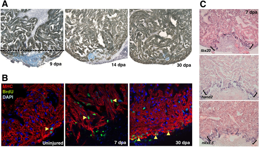
(a) Images of a regenerating zebrafish ventricle following 20% ventricular resection. Tissues were stained for myosin heavy chain to identify cardiac muscle (brown) and aniline blue to identify the fibrin clot (blue). By 7dpa, the wound is sealed by fibrin and is replaced by cardiac muscle by 30 dpa. (b) Proliferation, based on BrdU incorporation, is activated in cardiomyocytes by 7 dpa. The ventricular wall is restored by proliferation at the leading edge of the regenerating tissue. (c) Expression of embryonic heart field markers, hand2, nkx2.5, and tbx20 at the apical edge of the regenerate (brackets). Reproduced with permission from Refs. [7, 38].