
Figure 2. Participation of the epicardium during regeneration.
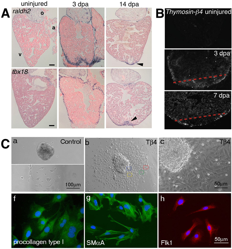
(a) Expression of the embryonic epicardial markers, raldh2 (top) and tbx18 (bottom) are induced in the adult epicardium after ventricular resection. Expression of both genes becomes localized to the wound by 14 dpa. The outflow tract (o), atrium (a), and ventricle (v) are labeled accordingly. (b) Radioactive in situ hybridization for Thymosin- 4 in sham operated control and amputated hearts 3 and 7 dpa. Expression of Thymosin- 4 is upregulated in regions surrounding the wound and the compact myocardium following injury. (c) Adult mouse heart explants cultured in vitro show increased migration of epicardial cells (confirmed by staining for epicardin – data not shown here) following treatment with Thymosin- 4. These cells then begin to express markers for smooth muscle cells (SM A – smooth muscle alpha-actin), fibroblasts (procollagen type I), and endothelial cells (Flk-1). Reproduced with permission from Refs. [38, 50, 52].