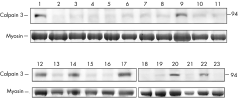
Figure 2 Screening of calpain‐3 autolytic activity in muscle biopsy specimens from patients with unclassified LGMD or hyperCKaemia. Lanes numbered 1–23 correspond to muscle samples from 23 different patients loaded after 5 min of incubation in saline solution, to promote calpain‐3 autolysis. The upper panel shows bands corresponding to the full‐length calpain‐3 protein at 94 kDa molecular weight, and the lower panel shows bands corresponding to myosin content in the post‐transfer Coomassie blue‐stained gel used to normalise the amount of calpain‐3 in each lane. In most muscle biopsy specimens analysed, there was full protein degradation, corresponding to normal function. Conversely, muscle samples numbered 1, 9, 12, 14, 17, 20 and 22 show the lack of protein degradation, resulting in a considerable amount of non‐degraded protein, which suggests the loss of normal autolytic function.

An official website of the United States government
Here's how you know
Official websites use .gov
A
.gov website belongs to an official
government organization in the United States.
Secure .gov websites use HTTPS
A lock (
) or https:// means you've safely
connected to the .gov website. Share sensitive
information only on official, secure websites.
