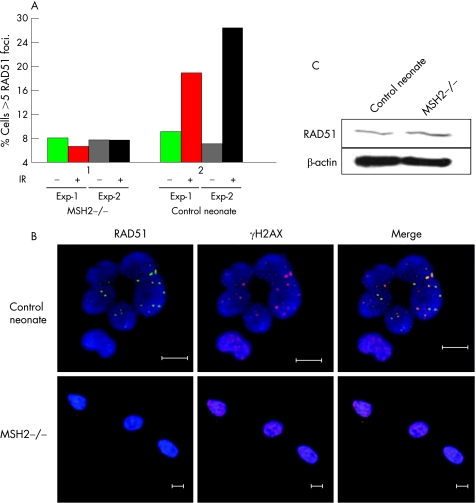
Figure 3 Reduced DNA repair intermediates in MSH2‐deficient cells. (A) Percentage of untreated (grey) and irradiated (10 Gy) (black) MSH2‐deficient and age‐matched control fibroblasts exhibiting >5 RAD51 foci; 100 γH2AX positive nuclei per experiment were evaluated for the presence of RAD51 foci. (B) Representative immunohistochemical staining of MSH2‐deficient and control cells. The presence of γH2AX foci (red) was used as a marker of double‐stranded DNA damage. Immunostaining of RAD51 (ab213, Abcam) is shown in green and nuclei labelled with Hoechst stain are blue. Bar: 10 μm. (C) Immunoblot of RAD51 protein expression in MSH2‐deficient and age‐matched control fibroblasts. β‐actin loading control is shown.

An official website of the United States government
Here's how you know
Official websites use .gov
A
.gov website belongs to an official
government organization in the United States.
Secure .gov websites use HTTPS
A lock (
) or https:// means you've safely
connected to the .gov website. Share sensitive
information only on official, secure websites.
