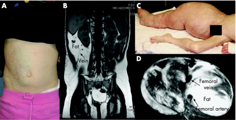
Figure 1 (A, B) Patient 8, a 5‐year‐old girl, presented with (A) right flank/abdominal mass with pinkish‐blue skin discoloration; (B) T1‐weighted MRI of this patient's abdomen showed a fast‐flow lesion, dilated draining vein (arrowed) and excessive fat. (C, D) Patient 25 had (C) an arteriolovenular anomaly and epidermoid naevus involving right lower limb, scrotum and penis; (D) T1‐weighted MRI of this patient's right thigh. Parental/guardian informed consent was obtained for publication of these figures.

An official website of the United States government
Here's how you know
Official websites use .gov
A
.gov website belongs to an official
government organization in the United States.
Secure .gov websites use HTTPS
A lock (
) or https:// means you've safely
connected to the .gov website. Share sensitive
information only on official, secure websites.
