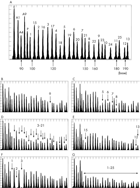
Figure 1 Multiplex ligation‐dependent probe amplification (MLPA) analysis of a control subject and patients with non‐ketotic hyperglycinaemia (NKH) with neonatal onset. A representative MLPA chromatogram of a control participant (A). The five control peaks include EXT2 exon 13 (E), AMT exons 1, 4 and 9 (A1, A4, A9), and GLDCP (P). The number on each peak indicates the exon number of the GLDC gene. MLPA probe for GLDC exon 14 was not used in this assay. MLPA analysis of patients with NKH: homozygotic deletion of exon 9 (B), heterozygotic deletion of exons 5–8 (C), heterozygotic deletion of exons 3–21 (D), heterozygotic deletion of exons 12–15 (E), homozygotic deletion of exons 1–3 (F) and heterozygotic deletion of all 25 GLDC exons (G).

An official website of the United States government
Here's how you know
Official websites use .gov
A
.gov website belongs to an official
government organization in the United States.
Secure .gov websites use HTTPS
A lock (
) or https:// means you've safely
connected to the .gov website. Share sensitive
information only on official, secure websites.
