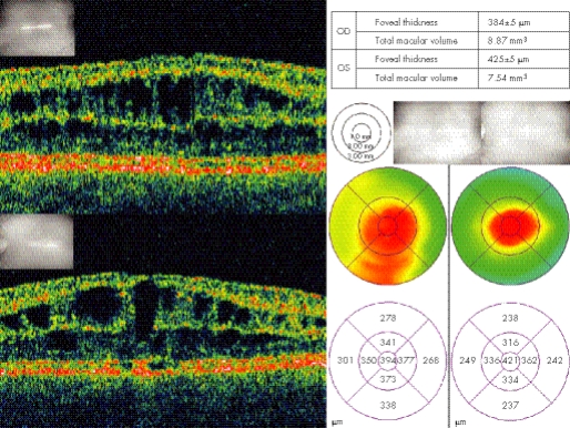
Figure 3 Optical coherence tomography images of the macular region from the same patient as fig 1. Top and bottom left showing schisis through multiple inner layers of the retina in right and left eyes, respectively. Macular thickness maps showing central macular thickening diffuse in right and more focal in the left eye.

An official website of the United States government
Here's how you know
Official websites use .gov
A
.gov website belongs to an official
government organization in the United States.
Secure .gov websites use HTTPS
A lock (
) or https:// means you've safely
connected to the .gov website. Share sensitive
information only on official, secure websites.
